The Role of Radiographic Evidence in Proving Causation for Extraction Claims

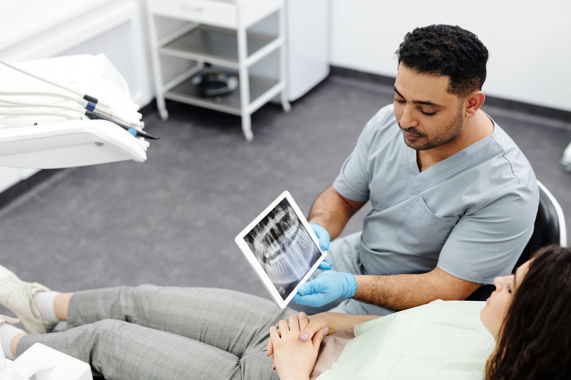
In clinical negligence litigation involving tooth extraction, the presence and quality of radiographic evidence often serve as an objective reference point when evaluating the standard of care and the nature of the underlying dental pathology. Whether a claim involves an allegation of wrong-tooth extraction, the management of periodontal disease, or injury to anatomical structures such as the inferior alveolar nerve, the court relies on the dental expert witness to interpret serial imaging. For instructing solicitors and barristers, understanding how these diagnostic records substantiate or rebut arguments of causation is central to the litigation strategy.
The Diagnostic Foundation of Extraction Claims
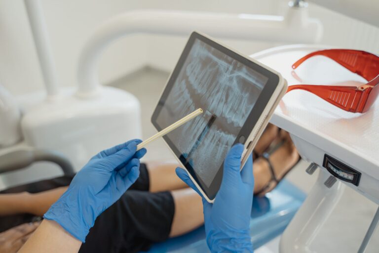
Radiographs provide a record of the clinical state of the dentition at the point of treatment. In cases involving alleged negligence, the court assesses whether the radiographic justification for extraction was sufficient and whether the procedure was performed with reasonable care. A specialist in managing complex restorative dental care often evaluates apical health, periodontal bone levels, and the proximity of tooth roots to adjacent structures. Where a clinician decides to extract a tooth, they generally rely upon a combination of clinical assessment and diagnostic imaging. The absence of a pre-operative radiograph where one is clinically indicated is a matter for expert evaluation regarding whether the clinician’s diagnostic approach met the standard of a responsible body of professional opinion.
Evidential Role in Breach of Duty and Causation
In assessing breach, the expert witness examines whether the clinician acted in accordance with a responsible body of professional opinion, as established in Bolam v Friern Hospital Management Committee [1957], and whether that opinion possesses a logical basis under Bolitho v City and Hackney Health Authority [1998]. Radiographic evidence often provides the data required to scrutinise these standards:
- Wrong-tooth extraction: Verification of the correct surgical site involves comparing pre-operative radiographs against clinical notes to ensure the tooth identified on the imaging matches the treatment record.
- Periodontal disease: Where a claimant alleges that a tooth was extracted prematurely due to undiagnosed or untreated periodontitis, the expert may scrutinise serial radiographs to determine the rate of bone loss and the clinical prognosis at the material time.
- Nerve injury: In cases involving the extraction of impacted third molars, the expert assesses whether the proximity of the roots to the inferior alveolar nerve was evaluated using panoramic radiographs or, where clinically indicated, cone-beam computed tomography (CBCT) imaging.
Causation requires a focus on whether, but for the breach, the harm would have been avoided. An expert report must establish that the radiographic findings would have altered the clinical management, rather than merely identifying a diagnostic oversight. For instance, if a radiograph reveals significant bone loss, the expert assesses whether a reasonable dentist would have nonetheless attempted to save the tooth, or if extraction was the likely clinical trajectory regardless of the quality of the baseline diagnostic imaging.
Common Pitfalls in Medico-Legal Casework
Solicitors should be aware of potential difficulties when managing dental extraction claims. A common complication involves over-relying on a single radiograph without context from the patient’s longitudinal history. Dental conditions are often progressive, and the expert’s duty under CPR 35.3 requires an objective assessment of the patient’s records as a whole. Furthermore, there is a distinction between a breach of duty and an inherent clinical risk. The doctrine of informed consent, governed by Montgomery v Lanarkshire Health Board [2015], requires that patients are warned of material risks. In extraction cases, the presence of anatomical landmarks on a radiograph that indicate a heightened risk of nerve damage is a factor in determining whether the patient was adequately informed.
Practical Guidance for Instructing Solicitors
To ensure a robust expert report, solicitors should obtain all relevant clinical records and imaging, including digital files in their original format where possible, as these may contain metadata useful for verifying dates. When preparing instructions, consider the following:
- Ensure the expert is matched to the specific sub-discipline. A general dental practitioner may have appropriate experience for routine extractions, but cases involving complex nerve anatomy or pathology may necessitate an expert with specific oral and maxillofacial experience.
- Frame questions around the clinical decision-making process. Ask the expert to state whether the radiograph provided sufficient information to support the procedure and whether any identifiable pathology was missed due to clinical assessment.
- Distinguish between breach and causation. Ask the expert to address whether the outcome would have differed had the standard of care been met, accounting for the pre-existing health of the dentition.
Where experts are required to produce a joint statement under CPR 35.12, the focus remains on narrowing issues regarding the interpretation of the radiographs. This process, which is distinct from the instruction of a single joint expert under CPR 35.8, allows the court to understand where experts agree on clinical findings and where their professional interpretations diverge. A high-quality report will focus on these technical distinctions, providing the court with the necessary clarity to determine if the dental clinician’s actions fell below the accepted standard.
This article is for general informational purposes only and does not constitute legal or medical advice. Readers should seek appropriate professional guidance.